

Desserts
Easy Recipe for a Delicious Chocolate Pecan Pi...
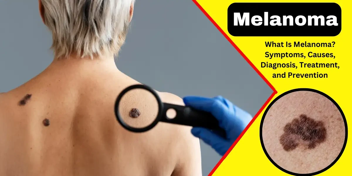
Melanoma is one of the most aggressive forms of skin cancer, originating in melanocytes—the cells that produce melanin, the pigment responsible for skin color. Although it accounts for only about 1% of all skin cancers, melanoma is responsible for the majority of skin cancer-related deaths due to its ability to metastasize (spread) quickly to other parts of the body. Early detection is critical, as survival rates are significantly higher when melanoma is treated before it advances.
Melanoma develops when melanocytes undergo genetic mutations, leading to uncontrolled cell growth. These mutations can be triggered by factors like ultraviolet (UV) radiation from the sun or tanning beds, genetic predisposition, or a weakened immune system. Melanoma often appears as an unusual mole or dark spot on the skin, but it can also arise in the eyes (ocular melanoma) or, rarely, in internal tissues like the digestive tract or mucous membranes.
Unlike basal cell carcinoma and squamous cell carcinoma—which are more common but less likely to spread—melanoma can invade nearby tissues and travel to distant organs, such as the lungs, liver, or brain. This aggressive behavior makes melanoma particularly dangerous. However, when detected early, it is highly treatable.
Key statistics highlight the importance of awareness:
Melanoma incidence has been steadily increasing over the past few decades, likely due to higher UV exposure and better detection methods.
The five-year survival rate for early-stage melanoma is over 99%, but it drops to around 30% if the cancer has spread to distant organs.
It is the leading cause of cancer death in young women (ages 25-29) and is also a significant concern for older adults.
Melanoma is not a single disease but rather a group of cancers with distinct characteristics. The main types include:
1. Superficial Spreading Melanoma: This is the most common type, accounting for about 70% of all melanoma cases. It typically begins as a flat or slightly raised lesion with irregular borders and varying colors (black, brown, red, or blue). It often develops on the trunk in men and the legs in women but can appear anywhere on the body. This type tends to grow horizontally across the skin before penetrating deeper layers.
2. Nodular Melanoma: Making up 15-30% of cases, nodular melanoma is more aggressive. It appears as a raised, firm bump, usually black or red, and grows rapidly—sometimes within weeks. Unlike superficial spreading melanoma, it does not have a prolonged horizontal growth phase, making it harder to catch early.
3. Lentigo Maligna Melanoma: This type is most common in older adults, particularly those with a history of chronic sun exposure. It begins as a flat, tan or brown patch (lentigo maligna) and slowly evolves into an invasive melanoma over years. It is frequently found on the face, neck, and arms.
4. Acral Lentiginous Melanoma: Although rare overall, this is the most common melanoma in people with darker skin tones. It appears on palms, soles, or under nails (subungual melanoma). Because it occurs in less sun-exposed areas, it is often diagnosed late, leading to poorer outcomes.
5. Amelanotic Melanoma: This rare form lacks pigment, making it difficult to detect. It may appear as a pink, red, or flesh-colored lesion, often mistaken for a benign growth. Due to its atypical appearance, diagnosis is frequently delayed.
Melanoma, the most serious type of skin cancer, often develops in existing moles or appears as a new, unusual growth on the skin. Recognizing the early signs of melanoma is crucial for early detection and successful treatment. The most common warning signs can be remembered using the ABCDE rule, but other symptoms, such as changes in sensation or texture, may also indicate melanoma. Below is a detailed explanation of the key symptoms and signs to watch for.
One of the primary indicators of melanoma is asymmetry. Unlike common moles, which are usually symmetrical (meaning one half mirrors the other), melanomas often have an irregular shape. If you draw a line through the middle of a suspicious mole and the two halves do not match, it could be a warning sign. Asymmetry occurs because cancerous cells grow uncontrollably, leading to an uneven appearance.
Healthy moles typically have smooth, even borders. In contrast, melanomas often have uneven, scalloped, or notched edges. The borders may appear blurred or poorly defined, making the mole look like it is spreading outward. This irregularity is caused by the uncontrolled growth of abnormal pigment-producing cells.
Normal moles are usually a single shade of brown. Melanomas, however, often contain multiple colors or uneven color distribution. You may notice shades of black, brown, tan, red, white, or even blue within the same lesion. These color changes occur due to the irregular production of melanin (skin pigment) by cancerous cells.
While melanomas can be small when first detected, they are often larger than 6 millimeters (about the size of a pencil eraser). However, some melanomas may be smaller, so any mole that grows in size—regardless of its initial diameter—should be examined by a dermatologist.
Any change in a mole’s appearance over time is a significant warning sign. This includes changes in size, shape, color, elevation, or texture. A mole that becomes raised, develops a scaly surface, starts bleeding, or becomes itchy should be evaluated immediately. Melanomas evolve because cancer cells continue to mutate and grow unpredictably.
While most moles are painless, melanomas may cause itching, tenderness, or even pain. If a mole becomes irritated, starts bleeding without injury, or forms a crust, it could indicate cancerous changes. These symptoms occur due to inflammation and the rapid growth of abnormal cells.
Melanomas may sometimes resemble non-healing sores or ulcers. If a spot or mole repeatedly scabs over but does not heal within a few weeks, it could be a sign of skin cancer. This happens because cancer disrupts the skin’s normal healing process.
In some cases, the pigment from a melanoma may spread into the surrounding skin, causing a "halo" or irregular darkening around the mole. This occurs when cancerous cells invade nearby tissues, leading to discoloration beyond the original lesion.
If you notice any of these signs, consult a dermatologist immediately. Early detection greatly improves treatment outcomes. Regular skin self-exams and annual dermatological check-ups are essential, especially for individuals with a family history of melanoma, fair skin, or excessive sun exposure. Remember, not all melanomas follow the ABCDE rule, so any new, changing, or unusual skin growth should be evaluated by a professional.
Melanoma is an aggressive form of skin cancer that arises from melanocytes, the cells responsible for producing melanin, the pigment that gives skin its color. While melanoma accounts for only about 1% of all skin cancers, it is responsible for the majority of skin cancer-related deaths due to its potential to metastasize if not detected early. Understanding the causes and risk factors of melanoma is essential for prevention and early intervention.
The most well-established cause of melanoma is exposure to ultraviolet (UV) radiation, primarily from the sun but also from artificial sources like tanning beds. UV radiation damages the DNA in skin cells, leading to mutations that can cause cells to grow uncontrollably and form cancerous tumors. There are two main types of UV rays that contribute to melanoma:
UVA rays penetrate deep into the skin and contribute to skin aging and long-term DNA damage.
UVB rays are primarily responsible for sunburn and play a key role in the development of skin cancers.
Both types of UV radiation are harmful, and repeated exposure—especially without adequate protection—increases melanoma risk. Studies have shown that individuals who use tanning beds before the age of 35 increase their melanoma risk by 75%, highlighting the dangers of artificial UV exposure.
People with fair skin, light hair (blonde or red), and light-colored eyes (blue or green) are at a significantly higher risk of developing melanoma. This is because their skin produces less melanin, the pigment that provides some natural protection against UV damage. Those with freckles or a tendency to burn rather than tan are also at increased risk.
However, melanoma can occur in individuals with darker skin tones as well. While they have more natural protection due to higher melanin levels, melanomas in these individuals often develop in less sun-exposed areas, such as the palms, soles, or under the nails (acral lentiginous melanoma), making them harder to detect early.
One of the most significant risk factors for melanoma is a history of severe, blistering sunburns, particularly during childhood or adolescence. Research indicates that five or more blistering sunburns in youth can double the lifetime risk of melanoma. This is because intense, intermittent UV exposure (such as getting sunburned on vacation) is more damaging than consistent, moderate exposure.
The presence of multiple moles (more than 50) increases melanoma risk. Additionally, individuals with atypical moles (dysplastic nevi)—which are larger than normal moles, have irregular borders, and varied colors—are at higher risk. Dysplastic nevi can sometimes transform into melanoma, and those with a family history of both dysplastic nevi and melanoma have an even greater risk.
About 10% of melanomas are hereditary, linked to specific gene mutations passed down in families. Key genetic mutations associated with melanoma include:
CDKN2A (p16) mutation – The most common hereditary melanoma gene, significantly increasing lifetime risk.
CDK4 mutation – Another gene linked to familial melanoma.
BRCA2 and BAP1 mutations – Also associated with higher melanoma risk.
If a first-degree relative (parent, sibling, or child) has had melanoma, an individual’s risk increases two to three times. Genetic testing may be recommended for families with a strong history of melanoma.
A compromised immune system reduces the body’s ability to detect and destroy cancerous cells, increasing melanoma risk. Conditions and factors that weaken immunity include:
Organ transplant recipients (due to immunosuppressive drugs).
HIV/AIDS patients.
Chronic lymphocytic leukemia (CLL) or lymphoma patients.
Long-term use of immunosuppressive medications (e.g., for autoimmune diseases).
While melanoma can occur at any age, risk increases with age, particularly after 50. However, it is one of the most common cancers in young adults (especially women under 30) due to tanning bed use and sun exposure habits.
Men are more likely to develop melanoma on the torso, head, or neck, while women more commonly develop it on the legs. Overall, men have a higher mortality rate from melanoma, possibly due to later detection.
Xeroderma pigmentosum (XP) is an inherited disorder that impairs the skin’s ability to repair UV-induced DNA damage. Individuals with XP have an extremely high risk of developing melanoma and other skin cancers, often at a very young age. Strict sun avoidance is critical for these patients.
Living near the equator – Higher UV intensity increases melanoma rates.
High-altitude locations – UV exposure is stronger at higher elevations.
Occupational sun exposure – Outdoor workers (farmers, construction workers) face higher cumulative UV damage.
Previous melanoma or non-melanoma skin cancer – Increases the risk of a new melanoma.
Parkinson’s disease – Some studies suggest a link, possibly due to shared genetic factors.
Chemical exposures – Certain industrial chemicals (arsenic, coal tar) may contribute.
Melanoma, the most serious type of skin cancer, is classified into stages based on tumor thickness, ulceration, lymph node involvement, and metastasis. The staging system helps determine the severity of the cancer and guides treatment decisions. The American Joint Committee on Cancer (AJCC) TNM system is commonly used, dividing melanoma into five main stages (0 through IV).
Stage 0, also called melanoma in situ, is the earliest and most treatable form. At this stage, cancerous cells are confined to the epidermis (the top layer of skin) and have not invaded deeper layers or spread to other parts of the body. Since the cancer is localized, surgical removal is usually curative, with a very high survival rate. Regular skin checks are still recommended to monitor for recurrence or new melanomas.
Stage I melanoma is divided into IA and IB based on tumor thickness and ulceration (whether the skin over the tumor is broken). In Stage IA, the tumor is less than 1 mm thick without ulceration. In Stage IB, the tumor is either:
Less than 1 mm thick with ulceration, or
Between 1 and 2 mm thick without ulceration.
At this stage, the cancer has not spread to lymph nodes or distant sites. Treatment typically involves surgical excision with wide margins, and the prognosis is excellent, with a 5-year survival rate of over 90%.
Stage II melanoma indicates a thicker tumor with a higher risk of spreading but no evidence of lymph node or distant metastasis. It is subdivided into:
IIA: Tumor is 1–2 mm thick with ulceration or 2–4 mm thick without ulceration.
IIB: Tumor is 2–4 mm thick with ulceration or over 4 mm thick without ulceration.
IIC: Tumor is over 4 mm thick with ulceration.
Because of the increased thickness and possible ulceration, Stage II melanoma has a higher chance of recurrence. Treatment involves surgical removal, and in some cases, sentinel lymph node biopsy (SLNB) may be recommended. Adjuvant therapies (such as immunotherapy or targeted therapy) might be considered for high-risk cases.
Stage III melanoma means the cancer has spread to nearby lymph nodes or skin but not to distant organs. It is further categorized based on the number of affected lymph nodes and whether the cancer has spread to nearby skin (satellite or in-transit metastases).
IIIA: Minimal lymph node involvement (microscopic spread detected via SLNB).
IIIB: One to three affected lymph nodes or local skin metastases.
IIIC: Four or more affected lymph nodes, matted lymph nodes, or extensive local spread.
Treatment involves surgical removal of the tumor and affected lymph nodes, followed by adjuvant therapy (immunotherapy or targeted therapy). The 5-year survival rate varies (40–70%) depending on the extent of lymph node involvement.
Stage IV is the most advanced stage, where melanoma has spread to distant organs such as the lungs, liver, brain, or bones. Symptoms depend on the affected organs and may include fatigue, weight loss, breathing difficulties, or neurological issues.
Treatment focuses on controlling the disease and improving quality of life. Options include:
Immunotherapy (e.g., checkpoint inhibitors like pembrolizumab)
Targeted therapy (for BRAF-mutant melanomas)
Radiation therapy (for symptom relief)
Chemotherapy (less commonly used now)
The prognosis for Stage IV melanoma has improved with newer treatments, but survival rates remain lower, with a 5-year survival rate of about 15–20%.
Melanoma staging is crucial for determining treatment and predicting outcomes. Early detection (Stages 0–II) leads to high cure rates, while advanced stages (III–IV) require aggressive therapies. Regular skin checks, sun protection, and prompt medical attention for suspicious moles are key to preventing and detecting melanoma early.
1. Clinical Examination: The diagnosis of melanoma typically begins with a thorough clinical examination by a dermatologist or healthcare provider. During this assessment, the doctor examines suspicious skin lesions using the "ABCDE" rule—checking for Asymmetry, irregular Borders, uneven Color, large Diameter (greater than 6mm), and Evolution (changes in size, shape, or texture). Additionally, the physician may inquire about the patient’s medical history, including sun exposure, family history of skin cancer, and any previous skin abnormalities. If a lesion appears concerning, further diagnostic tests, such as a biopsy, are recommended.
2. Dermoscopy: Dermoscopy, also known as dermatoscopy, is a non-invasive imaging technique that allows doctors to examine skin lesions more closely. Using a handheld device called a dermatoscope, the physician magnifies the lesion and observes its structural patterns, colors, and blood vessels, which are not visible to the naked eye. This method improves diagnostic accuracy by distinguishing between benign moles and malignant melanomas. In some cases, digital dermoscopy is used to track changes in lesions over time, aiding in early detection.
3. Biopsy and Histopathological Analysis: If a lesion is highly suspicious for melanoma, a biopsy is performed to confirm the diagnosis. The most common biopsy techniques include excisional biopsy (removing the entire lesion), incisional biopsy (removing a portion), or punch biopsy (using a circular tool to extract a tissue sample). The collected tissue is sent to a pathology lab, where a histopathological examination is conducted. A pathologist examines the sample under a microscope to assess the Breslow thickness, ulceration, mitotic rate, and other features that determine the stage and aggressiveness of the melanoma.
4. Molecular and Genetic Testing: In advanced or high-risk melanomas, molecular and genetic testing may be performed to identify specific mutations, such as BRAF, NRAS, or c-KIT. These tests help guide targeted therapy and immunotherapy options. For example, patients with a BRAF V600E mutation may benefit from BRAF inhibitor drugs like vemurafenib or dabrafenib. Additionally, genetic profiling can assess the risk of metastasis and aid in personalized treatment planning.
5. Imaging Tests for Staging: Once melanoma is confirmed, imaging tests such as CT scans, MRI, PET scans, or ultrasound may be used to determine if the cancer has spread (metastasized) to lymph nodes or other organs. Sentinel lymph node biopsy (SLNB) is often performed in cases where melanoma is deeper than 1mm to check for microscopic spread. Staging (from Stage 0 to Stage IV) is crucial for determining prognosis and selecting the most effective treatment strategy.
Melanoma is an aggressive form of skin cancer that develops when melanocytes undergo malignant transformation due to DNA damage, often from UV radiation exposure. While early detection leads to high cure rates, advanced melanoma requires a multifaceted treatment approach. This article explores current treatment modalities, including surgical options, immunotherapy, targeted therapy, chemotherapy, radiation, and emerging therapies, providing an in-depth understanding of melanoma management.
Surgery is the primary treatment for localized melanoma. The extent of excision depends on tumor thickness (Breslow depth) and location.
Wide Local Excision: The standard approach involves removing the tumor with a margin of healthy tissue (0.5–2 cm) to ensure complete eradication.
Sentinel Lymph Node Biopsy (SLNB): Used for tumors deeper than 0.8 mm to detect microscopic spread. If positive, a complete lymph node dissection may follow.
Mohs Surgery: For melanomas in cosmetically sensitive areas (e.g., face), this technique preserves healthy tissue while ensuring complete removal.
Surgery may also be used palliatively in metastatic cases to relieve symptoms.
Immunotherapy has revolutionized melanoma treatment by enhancing the body’s immune system to recognize and attack cancer cells. The most widely used immunotherapies are immune checkpoint inhibitors, which block proteins that prevent immune cells from attacking cancer.
PD-1 Inhibitors (Pembrolizumab, Nivolumab): These drugs block the PD-1 receptor on T-cells, allowing them to recognize and destroy melanoma cells more effectively. Pembrolizumab (Keytruda) and nivolumab (Opdivo) are first-line treatments for advanced melanoma, significantly improving survival rates compared to older therapies.
CTLA-4 Inhibitor (Ipilimumab): Ipilimumab (Yervoy) targets the CTLA-4 protein, another immune checkpoint that suppresses T-cell activity. While effective, it has a higher risk of severe immune-related side effects, such as colitis, hepatitis, and endocrine disorders.
Combination Therapy (Nivolumab + Ipilimumab): Combining PD-1 and CTLA-4 inhibitors increases response rates and long-term survival but also raises the risk of toxicity. Patients on this regimen require close monitoring for autoimmune complications.
Cytokine Therapy (Interleukin-2, Interferon-alpha): These older treatments stimulate the immune system but are less commonly used today due to severe side effects (e.g., high fever, capillary leak syndrome) and the superior efficacy of checkpoint inhibitors.
Targeted therapies are designed for melanomas with specific genetic mutations, such as BRAF, NRAS, or KIT. These drugs block the signaling pathways that drive cancer growth.
BRAF Inhibitors (Vemurafenib, Dabrafenib, Encorafenib): About 50% of melanomas have a BRAF V600E or V600K mutation, making them susceptible to BRAF inhibitors. These drugs (e.g., vemurafenib/Zelboraf, dabrafenib/Tafinlar) shrink tumors rapidly but often lead to resistance within months.
MEK Inhibitors (Trametinib, Cobimetinib, Binimetinib): Since BRAF inhibitors alone can activate alternative pathways (like MEK), combining them with MEK inhibitors (e.g., trametinib/Mekinist) improves outcomes and delays resistance.
KIT Inhibitors (Imatinib, Nilotinib): Rare melanomas (e.g., mucosal or acral) may have KIT mutations, making them responsive to drugs like imatinib (Gleevec). However, these cases require genetic testing for confirmation.
Chemotherapy is now less commonly used for melanoma due to the success of immunotherapy and targeted drugs. However, it may still be an option for patients who do not respond to newer therapies.
Dacarbazine (DTIC): The most traditional chemotherapy for melanoma, with response rates of only 10-20%.
Temozolomide (Temodar): An oral alternative that can cross the blood-brain barrier, sometimes used for brain metastases.
Combination Chemotherapy (CVD: Cisplatin, Vinblastine, Dacarbazine): Rarely used due to toxicity and limited survival benefits.
Radiation is primarily used in melanoma for:
Adjuvant Therapy: After lymph node dissection to reduce recurrence risk.
Palliative Care: To relieve pain from bone metastases or control brain metastases.
Stereotactic Radiosurgery (SRS): A precise, high-dose radiation technique for isolated brain tumors.
T-Cell Therapy (TILs, CAR-T Cells)
Tumor-Infiltrating Lymphocytes (TILs): Immune cells extracted from the tumor, expanded in a lab, and reinfused to enhance anti-cancer activity.
CAR-T Therapy: Genetically engineered T-cells designed to target melanoma-specific antigens (still in clinical trials).
Oncolytic Virus Therapy (Talimogene Laherparepvec, T-VEC): A modified herpes virus (Imlygic) injected into tumors, causing cell destruction and immune activation.
BiTE Antibodies (Tebentafusp): A novel treatment for metastatic uveal melanoma, redirecting T-cells to attack cancer.
Each treatment has unique risks:
Immunotherapy: Autoimmune reactions (rash, colitis, thyroid dysfunction).
Targeted Therapy: Skin changes, fever, heart issues.
Chemotherapy: Fatigue, nausea, low blood counts.
Early intervention and supportive care are essential for maintaining quality of life.
Survival rates vary by stage:
Stage 0-II: Over 99% 5-year survival.
Stage III: 60-70% (depends on lymph node involvement).
Stage IV: 20-30% (depends on spread location).
Regular follow-ups are essential to monitor for recurrence.
Reduce risk with these strategies:
1. Limit Sun Exposure: One of the most effective ways to prevent melanoma is to limit direct exposure to ultraviolet (UV) rays from the sun. The sun's rays are strongest between 10 a.m. and 4 p.m., so it is advisable to seek shade during these hours. Prolonged sun exposure, especially without protection, increases the risk of skin damage and melanoma. Even on cloudy days, UV rays can penetrate the skin, so protective measures should be taken year-round.
2. Use Sunscreen Regularly: Applying a broad-spectrum sunscreen with an SPF of 30 or higher is essential in protecting the skin from harmful UV radiation. Sunscreen should be applied generously to all exposed skin at least 15–30 minutes before going outside and reapplied every two hours, or more frequently if swimming or sweating. Many people underestimate the amount needed—about one ounce (a shot glass full) is recommended for full-body coverage.
3. Wear Protective Clothing: Clothing provides a physical barrier against UV rays. Wearing long-sleeved shirts, wide-brimmed hats, and UV-blocking sunglasses helps shield the skin and eyes from sun damage. Some clothing is specially designed with ultraviolet protection factor (UPF) ratings, which indicate how effectively the fabric blocks UV radiation. Darker, tightly woven fabrics generally offer better protection than light, thin materials.
4. Avoid Tanning Beds: Tanning beds emit concentrated UV radiation, which significantly increases the risk of melanoma and other skin cancers. Studies have shown that using tanning beds before the age of 35 raises melanoma risk by nearly 60%. Instead of artificial tanning, safer alternatives like self-tanning lotions or spray tans can provide a sun-kissed look without the harmful effects of UV exposure.
5. Perform Regular Skin Self-Exams: Early detection of melanoma greatly improves treatment outcomes. Individuals should examine their skin monthly for new or changing moles, freckles, or spots. The ABCDE rule can help identify suspicious growths: Asymmetry, irregular Borders, uneven Color, large Diameter (greater than 6mm), and Evolving (changing in size, shape, or color). Any concerning changes should be evaluated by a dermatologist.
6. Get Professional Skin Checks: In addition to self-exams, annual skin checks by a dermatologist are recommended, especially for those with a family history of melanoma, fair skin, or numerous moles. Dermatologists can detect early signs of melanoma that may be missed during self-examinations. People at higher risk may need more frequent screenings.
7. Protect Children from Sun Damage: Childhood sunburns significantly increase the risk of melanoma later in life. Parents should ensure children wear sunscreen, protective clothing, and hats when outdoors. Infants under six months should be kept in the shade, as their skin is highly sensitive to UV damage. Teaching sun-safe habits early helps establish lifelong protective behaviors.
Melanoma, the most aggressive form of skin cancer, can lead to severe complications if not detected and treated early. These complications arise when the cancer spreads (metastasizes) to other parts of the body or when treatments cause significant side effects. Below are the key complications associated with melanoma:
One of the most serious complications of melanoma is its ability to metastasize to distant organs. Initially, melanoma may appear as a mole or dark spot on the skin, but if left untreated, it can invade deeper layers of the skin and enter the lymphatic system or bloodstream. Common sites of metastasis include the lungs, liver, brain, bones, and gastrointestinal tract. Once melanoma spreads, it becomes much more difficult to treat and significantly reduces survival rates. Symptoms depend on the affected organ—for example, shortness of breath (lungs), headaches (brain), or bone pain (skeletal system).
Melanoma often spreads first to nearby lymph nodes, causing them to swell and become firm or painful. Lymph node involvement indicates a more advanced stage of cancer and may require surgical removal (lymphadenectomy) or additional treatments like immunotherapy or radiation. If cancer cells block lymphatic vessels, it can lead to lymphedema—a condition where fluid builds up, causing swelling, usually in the arms or legs. This can result in discomfort, limited mobility, and an increased risk of infections.
Even after successful treatment, melanoma can return, either at the original site or in other parts of the body. The risk of recurrence depends on factors such as tumor thickness, ulceration, and whether the cancer had spread initially. Patients who have had melanoma require lifelong follow-ups with regular skin exams and imaging tests to detect any new or returning tumors early. Recurrent melanoma is often more aggressive and may require stronger treatments, such as targeted therapy or chemotherapy.
Treatments for melanoma, including surgery, immunotherapy, targeted therapy, radiation, and chemotherapy, can cause significant side effects. Surgery may lead to scarring, nerve damage, or infections. Immunotherapy, while effective, can trigger autoimmune reactions, causing fatigue, skin rashes, or organ inflammation. Targeted therapies may result in high blood pressure, liver problems, or heart complications. Radiation can cause skin burns and long-term tissue damage. Managing these side effects is crucial to maintaining a patient’s quality of life during and after treatment.
A melanoma diagnosis can cause anxiety, depression, and emotional distress, especially in advanced cases. Patients may struggle with fear of recurrence, changes in appearance due to surgery, or the physical toll of treatment. Support groups, counseling, and mental health care are essential in helping patients and their families cope with the emotional challenges of melanoma.
Melanoma is a serious but often preventable cancer. Knowing the warning signs, risk factors, and prevention methods can make a life-saving difference. If you notice any suspicious skin changes, don’t wait—consult a dermatologist immediately.
Stay sun-smart, check your skin regularly, and prioritize early detection for the best outcomes.
The ABCDE rule highlights key warning signs:
Asymmetry: One half of the mole looks different.
Border irregularity: Ragged or blurred edges.
Color variation: Multiple shades (black, brown, red, white).
Diameter: Larger than 6mm (but early melanomas can be smaller).
Evolving: Changes in size, shape, or texture.
Bonus sign: A new mole in adulthood or a sore that won’t heal.
Early-stage (0–II): Nearly 100% curable with surgical removal.
Advanced (III–IV): Not always curable but treatable. Survival rates drop to ~20–30% if it spreads to distant organs.
Stage 0: Melanoma in situ (top skin layer only).
Stage I/II: Localized, increasing thickness (up to 4mm+).
Stage III: Spread to nearby lymph nodes.
Stage IV: Metastasized to distant organs (lungs, liver, brain).
Primary cause: DNA damage in melanocytes (often from UV radiation).
Other triggers: Genetic mutations (e.g., BRAF), family history, or immune suppression.
Yes, if caught early (Stage 0–II).
Advanced melanoma: Managed with immunotherapy, targeted therapy, or surgery, but not always curable.
High-risk groups include:
People with fair skin, light eyes, or red/blonde hair.
Those with many moles or atypical moles.
Individuals with a history of sunburns or tanning bed use.
People with family history of melanoma.
Immunosuppressed individuals (e.g., organ transplant recipients).
Early melanoma: Usually not painful (may itch or bleed).
Advanced melanoma: Can cause pain if it spreads to bones/nerves.
Sun protection: SPF 30+, hats, shade.
Avoid tanning beds (75% higher risk).
Regular skin checks (self-exams + dermatologist visits).
Most common in fair-skinned adults over 50, but young adults (especially women 25–29) are at rising risk.
Darker-skinned individuals: Higher risk of acral lentiginous melanoma (palms, soles, nails).
Not all moles are melanoma, but melanoma often looks like an irregular mole.
30–50% of melanomas arise from existing moles; the rest develop on normal skin.
Typically spreads to:
Lymph nodes (closest to the tumor).
Lungs.
Liver.
Brain.
Bones.
The highest-risk patient has:
Fair skin + history of sunburns.
Many atypical moles.
Family history of melanoma.
Weakened immune system.